
Advantages of halogen over L.E.D. illumination in darkfield microscopy
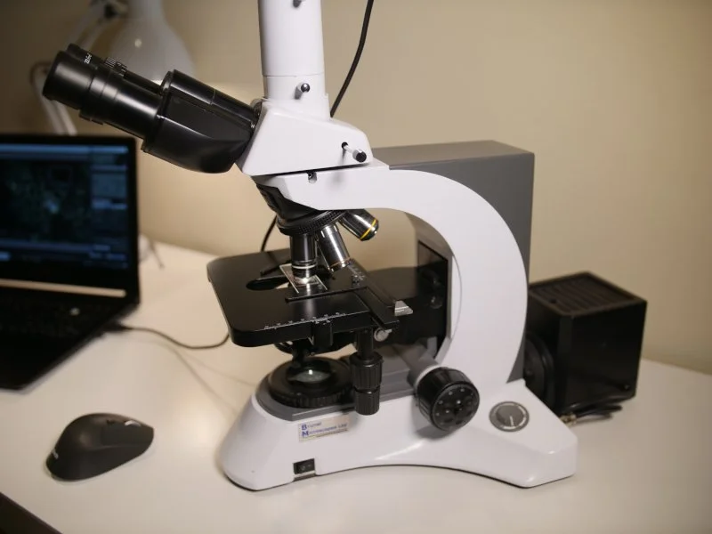
Darkfield microscope with a halogen bulb mounted in an external light box
Modern darkfield microscopes are supplied with either halogen or L.E.D. light sources to illuminate the sample. A common question asked when students are looking to purchase a darkfield microscope is “Which is better - a halogen or L.E.D. light source?”
To answer this question, we need to understand the importance of light in darkfield microscopy.
In brightfield microscopy the light in the microscope shines directly through the sample and into the objectives, which are the lenses that magnify the sample. This works well for samples that have non-transparent or opaque objects in them so that light bounces off the objects. Then, as you look through the microscope’s binoculars you will see the magnified sample brightly lit.
In darkfield microscopy a condenser is placed between the light source and the sample and this condenser disrupts the flow of light. The light is blocked and diverted so that it enters the sample at an angle.
When looking at blood under a microscope this is crucial because live blood is almost transparent in brightfield and you cannot see the details of components such as cytoplasmic contents, nuclei, membranes and plasma components.
This angled light flows through the sample and bounces off the fine components in blood, making what was invisible in brightfield, suddenly completely illuminated.
The darkfield condenser’s scattering of light inevitably means it will “lose” light which is why it is CRUCIAL to have a strong light source to begin with.
In summary the two main issues for Live Blood Analysis are:
A Live Blood sample is virtually invisible using brightfield microscopy
To see Live Blood in darkfield microscopy you need a strong light!
Which is better for darkfield, L.E.D. or halogen?
First you need to make sure that whichever light source you use, it is strong enough to illuminate the blood in darkfield.
You can get some detail with less wattage in both L.E.D.and halogen, but as professional analysts it is crucial that we can see as much detail as possible, particularly all the fine components in the blood.
Halogen vs L.E.D.
Here are several points to show the difference between using halogen and L.E.D. light sources in darkfield microscopy:
Halogen is rare - Nearly all darkfield microscopes now use L.E.D. and so finding a model that uses halogen is rare. This is mainly due to shifts in industry where L.E.D. is considered more energy efficient.
Halogen is preferred for darkfield - Interestingly in darkfield, halogen is still the preferred lighting method. It emits a smooth, continuous spectrum of light and a natural rich colour reproduction, which really helps with identification of different cellular components. L.E.D. has a more blue/white colour spectrum and it is weaker in intensity.
Halogen vs L.E.D. in brightness – In order to match a 100w halogen bulb in the level of light it emits, the L.E.D. bulb needs to be a 10-15w L.E.D. It is very rare to find a microscope that uses this strong an L.E.D. bulb.
The exception to this is the Euromex iScope L.E.D. microscope that we have trialled and we recommend. It has 2 light sources to get over this issue of needing a strong light for darkfield. The darkfield condenser has an integrated L.E.D. 3w bulb in it which means the light source is extremely close to the sample and illuminates it greatly. There is a second 3W L.E.D. bulb in the foot of the microscope for brightfield.
Heat from the bulb can affect the sample - Halogen bulbs give off more heat than L.E.D. bulbs and it is for this reason that the Brunel Haemascope’s 100w halogen bulb is housed in an external ventilated light box. The L.E.D. in the Euromex iScope is housed in the condenser, and although L.E.D. does not give off as much heat as halogen, it is still emitting heat and as it is close to the sample it may heat it up slightly.
Natural Light - Halogen emits a light spectrum similar to natural sunlight in the ranges of 100 to 2500nm. It has a very high infrared component, but it covers all the visible light spectrum, allowing the sample to be naturally lit. Halogen is also a smooth, continuous spectrum of light.
Blue Light – L.E.D. light bulbs give off predominantly blue light, 450 – 490nm, which does not reflect true, natural light. L.E.D. bulbs have light spikes and they flicker and strobe at rates that are invisible to the naked eye, but our eyes and brains are still exposed to this flicker effect.

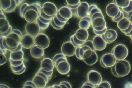
Live Blood sample taken using a halogen light source(illustration purposes only)
Live Blood sample taken using a L.E.D. light source(illustration purposes only)
So which is better?
Halogen, as long as it is bright enough [100w], and the heat from the bulb does not affect the sample, is superior for Live Blood Analysis using darkfield microscopy.
L.E.D. , as long as it is bright enough [3W housed in darkfield condenser or 12W in foot of microscope] is still suitable for Live Blood Analysis using darkfield microscopy (with the caveat that heat from the bulb may affect the sample).
BUT there is more………….. (see the section below)
For your interest
Here is a link to a blog article that I wrote where I show you the comparison between the Brunel 100w halogen microscope and a 3W LED microscope[with no light in the condenser], so you can see the difference.
Which is Healthier for your Eyes and Brain ?
The question of “which is better?” does not just relate to the image quality produced by these two light sources. There are also considerations for the health of microscopists using these devices, whether it is the impacts on physiology or sleep disruption.
Comparing halogen vs. L.E.D. light in terms of health impacts
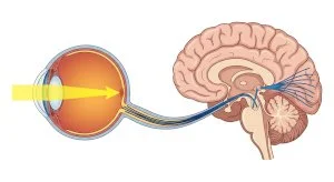
There is no doubt that busy microscopists and blood analysts will use a microscope often for hours every working day, over many years. Microscopes focus light up directly into their eyes, which are then connected to the brain.
We are already bombarded by non-native E.M.F.s (electro-magnetic fields)– mostly blue light spectrum. We literally overdose on it via phones, laptops, screens, lighting etc. These have been proven to impact our circadian rhythm (sleep cycle) and can have an impact on eye health.
Halogen bulbs emit in the same range as native E.M.F.s from natural sunlight. There really is no comparison from a health perspective when considering the eye and brain health of the user, and the growing field of biophysics and photo biomodulation prove this point.
Follow these links for further information.
https://www.chronobiologyinmedicine.org/journal/view.php?number=167&viewtype=pubreader
So whether it is their ability to generate a clear, detailed image, or to protect your health longterm as a microscopist, 100W halogen bulbs still represent the ‘gold-standard’ for darkfield microscopy when used for Live Blood Analysis.
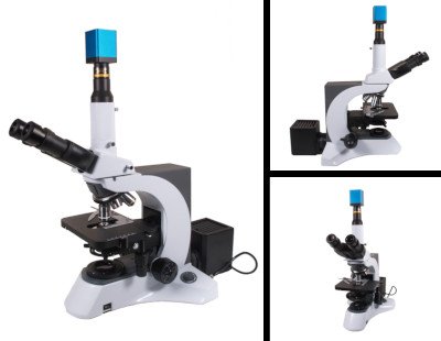
To learn more about our Brunel Haemascope 100W Halogen Darkfield Microscope & 4K Camera package click here.
Brunel Haemascope 100W Halogen Darkfield
If you would like to know more about Live and Dry Blood Analysis Training so that you can use it to assess your clients’ health then please follow this link.
You are also invited to join our Facebook Group ‘Learning Live Blood Analysis’ - a private group of students, qualified analysts and anyone interested in learning.
If you have any questions about this topic or LDBA in general please email Shirah directly at info@naturecureacademy.com or add a comment below!
